Description
Introduction. Tennis is a biomechanically demanding sport associated with specific injuries. The percentage of medial elbow injuries among elite tennis players compared to lateral elbow injuries is greater, imposing to uncover the mechanism of injury and the reason for this phenomenon. The goal of this study was to use image analysis recordings to measure the carrying angle of elite male tennis players during the forehand stroke, with the hypothesis that elite tennis players overstress their elbow in valgus over the physiological degree in the frontal plane just prior to ball contact on forehand groundstrokes.
Methods and Material. The carrying angle of male tennis players ranked in the top 25 position in the ATP ranking was measured on selected video frames with the elbow as close as possible to full extension just prior to the ball-racket contact in forehands. These frames were extracted from 306 videos professionally recorded for training purposes by a high-profile video analyst. All measures were conducted by three independent observers.
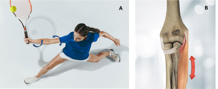
Results. Sixteen frames were finally included. The mean carrying angle was 11.50° ± 4.74°. The intraclass correlation coefficient value was 0.703, showing good reliability of the measurement technique. The measured carrying angle was lower than what has been observed in historical cohorts using comparable measurement methodology, suggesting a possible instant varus accommodation (DIVA) mechanism before hitting the ball (FIG 1).
Conclusions. The observed decrease in the carrying angle is a consequence of an increase in elbow flexion position dictated by the transition from a closed to open, semi open stances. As the elbow flexes during the preparation phase, it is less constrained by the olecranon and its fossa, increasing the strain on the medial collateral ligament and capsule structures. Moving toward full extension prior to the ball-racket contact, the elbow is dynamically stabilized by a contraction of the flexor muscles.